



AMOS1 .com's Store
easyid.live/biz/amos1 • Verified Store
Scan to open official EasyID digital store and view live catalog.
Share AMOS1 .com's Store
Share this store link with your friends and customers
AMOS1 .com's Store
Open Now • Open 24/7Verified Merchant
EasyID Certified Seller
Fast 24h Packing
Direct courier dispatch
Zero Escrow Direct
Direct merchant settlement
Store Listings (7)
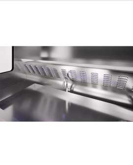
TST1200 Grossing Station for Histopathology
[a]https%3A%2F%2Fwww.amos-scientific.com%2Fproducts%2Ftst1200-grossing-station-for-histopathology.html[/a] <br>#automatic #tissue #processor #use <br>Our Pathology Grossing Stations are cutting-edge laboratory workstations designed for precise specimen examination and dissection. TST1200 histopathology grossing station featuring ergonomic design and advanced technology, they streamline the pathology workflow for accurate diagnostics. <br> <br>Description of TST1200 Grossing Station for Histopathology <br>stainless steel housing <br> <br>low exhaust system <br> <br>cold and hot water system <br> <br>air sterilizing lamp and lighting <br> <br>automatic induction type hand drier <br> <br>stainless steel table fitted with lateral spray system, washing sink and universal telescopic water tap <br> <br>pulverizer <br> <br>For pathology grossing station price and histopathology lab instruments, contact us.
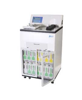
Tissue Processor
[a]https%3A%2F%2Fwww.amos-scientific.com%2Fproducts%2Ftissue-processor%2F[/a] <br>#automatic #tissue #processor <br>A Tissue Processor is a laboratory instrument used in medical and biological research settings for the processing of tissue specimens. It performs various steps such as fixation, dehydration, cleaning, and impregnation of tissue specimens. The Tissue Processor automates these processes, improving efficiency and accuracy. It plays a crucial role in pathology diagnosis, research, and education. <br> <br>Use of Automatic Tissue Processor <br>Advancements in Histology: The Essential Role of Automatic Tissue Processor <br> <br>In the realm of pathology and medical research, the Automatic tissue processing equipment(ATP) stands as a pivotal tool, revolutionizing the way tissue specimens are prepared for microscopic examination. This automated marvel plays a crucial role in histology laboratories, streamlining and enhancing the tissue processing workflow. <br> <br>Gone are the days of manual tissue processing, as ATPs have become indispensable in the modern laboratory setting. These devices are designed to efficiently and consistently process tissue specimens through a series of standardized steps, ensuring high-quality results. The automation provided by these processors not only saves time but also minimizes the risk of human error, promoting accuracy and reproducibility in histological analyses. <br> <br>One of the key features of Automatic Tissue Processors is their ability to handle multiple specimens simultaneously. This not only increases the efficiency of the laboratory but also allows for the processing of large batches of samples, crucial in studies involving extensive tissue analysis. The processors follow a pre-programmed sequence of steps, including fixation, dehydration, clearing, and infiltration with paraffin wax, ensuring that the tissues are adequately prepared for sectioning. <br> <br>The precise control over processing parameters provided by ATPs is another significant advantage. Researchers can tailor the settings based on the specific characteristics of different tissue types, optimizing the processing conditions for each sample. This level of customization contributes to the production of high-quality tissue sections, vital for accurate pathological diagnosis and research outcomes. <br> <br>Automatic Tissue Processor has become an indispensable tool in histology laboratories, transforming the landscape of tissue processing. Its automation, efficiency, and precision make it an essential asset for researchers and pathologists alike, facilitating. <br> <br>Components and Functions of a Tissue Processor <br>1. Control panel: The control panel allows the user to set and monitor processing parameters such as temperature, agitation, and processing time. It provides a user-friendly interface for programming and adjusting processing protocols. <br> <br>2. Processing chamber: The processing chamber is where the tissue specimens are placed. It is designed to hold various sizes and types of cassettes or containers, ensuring proper immersion in the processing reagents. The chamber should be resistant to chemical damage and easy to clean. <br> <br>3. Reagent containers and dispensers: Tissue processors have separate containers for each processing reagent, including fixatives, dehydrants, clearing agents, and embedding media. These containers are equipped with automatic dispensing mechanisms to ensure proper and consistent reagent delivery to the specimens. <br> <br>4. Temperature control system: Tissue processors have a temperature control system that maintains the desired processing temperature throughout the entire processing cycle. This ensures optimal tissue preservation and processing outcomes. <br> <br>5. Agitation mechanism: An agitation mechanism ensures thorough and uniform mixing of the specimens with the processing reagents. It can be in the form of oscillation, rocking, or rotation, depending on the specific tissue processor model. <br> <br>6. Fluid circulation system: A fluid circulation system facilitates the movement and exchange of processing reagents within the processing chamber, promoting consistent and efficient tissue processing. <br> <br>7. Safety features: Tissue processors should have safety features such as alarms, temperature monitoring, and emergency shut-off mechanisms to protect the specimens, operator, and instrument from potential risks or malfunctions. <br> <br>8. Drying system: Some tissue processors also have a built-in drying system to aid in the removal of residual fluids from the processed tissues before embedding. <br> <br>9. Programmable protocols: Tissue processors allow the user to program and store custom processing protocols for different types of specimens. This ensures optimal processing conditions and flexibility in adapting to specific requirements. <br> <br>Comparison Of Manual And Automated Tissue Processing Techniques <br>1. Workflow efficiency: Automated tissue processors offer significant time savings compared to manual processing. The automated system can handle multiple specimens simultaneously, reducing processing time and increasing laboratory productivity. Manual processing, on the other hand, is a time-consuming and labor-intensive process. <br> <br>2. Consistency and standardization: Automated tissue processors ensure consistent and standardized results due to their programmed protocols. This reduces the chances of errors and variations in processing times and reagent exposures. Manual processing, however, is subject to individual variances and human errors, leading to inconsistent results. <br> <br>3. Quality control: Automated tissue processors allow for precise control over processing conditions, such as temperature, agitation, and reagent volumes. This enhances the quality of the processed tissues and improves the overall reliability of the analysis. Manual processing is highly dependent on the skills and expertise of the individual operator, making quality control more challenging. <br> <br>4. Handling of large specimen volumes: Automated tissue processors are capable of handling a larger number of specimens simultaneously, making them suitable for high-volume laboratories. Manual processing is limited by the manual handling capability of the operator and may require more time and effort for large specimen volumes. <br> <br>5. Cost considerations: Automated tissue processors are generally more expensive to purchase and maintain compared to manual processing equipment. However, the increased workflow efficiency and consistency of results can lead to cost savings in the long run. <br> <br>In conclusion, automated tissue processing offers improved workflow efficiency, consistency, and standardization compared to manual techniques. However, the choice of processing method depends on the specific needs and resources of the laboratory. <br> <br>For automatic tissue processor price and histopathology laboratory apparatus, contact us.
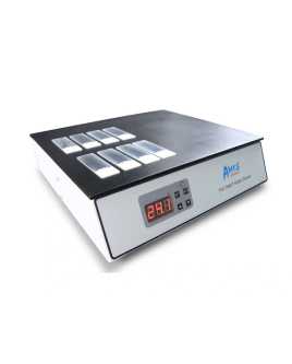
TEC2602 Slide Dryer
[a]https%3A%2F%2Fwww.amos-scientific.com%2Fproducts%2Ftec2602-slide-dryer.html[/a] <br>#dryer #slide <br>Experience efficient slide drying with our Pathological Slide Dryer. Designed for histology labs, it accelerates the drying process while maintaining sample integrity. This reliable instrument streamlines slide preparation for accurate pathological analysis and research applications. <br> <br>Description of TEC2602 dryer slide <br>Temperature can be set to user's favor. <br>Easy and reliable operation display. <br>Stand alone unit placed near microtome. <br>It suits all laboratories. <br> <br>Specification of TEC2602 Slide Dryer <br>Table top 324 x 272 mm <br>Temperature control accuracy ±1℃ <br>Temperature range ambient temperature ~90℃ <br>Capacity can load 40 slides simultaneously <br>Rated voltage 220V±10% <br>50/60 Hz / 110V±10% <br>50/60 Hz <br>Nominal power 350W <br>Outside dimensions(mm) 324(W) ×300(D)×85(H) <br>Weight 2.75kg <br> <br>There are many histology equipment manufacturers, but we are one of the best choices for you.
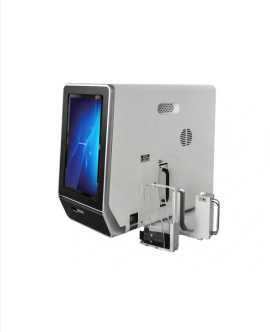
SM100 Slide Printer
[a]https%3A%2F%2Fwww.amos-scientific.com%2Fproducts%2Fsm100-slide-printer.html[/a] <br>#slide #label #printer <br>Our histology slide label printer is a lab essential for easy and efficient slide labeling. With precision and speed, it produces clear and permanent markings on glass slides, assisting organization and enhancing traceability in pathology and research settings. <br> <br>Characters of SM100 Slide Printer <br>Simple design, easy to use; <br> <br>High printing speed, 3-5 seconds per slide; <br> <br>Non-contact UV laser technology; <br> <br>Excellent ability to export characters, numbers, letters, 1D & 2D codes, graphics; <br> <br>Fully compatible with LIS/HIS system; <br> <br>11 inch LED touch screen via built-in software, also support to connect external PC; <br> <br>Built in bar-code scanner, and support to connect external scanners; <br> <br>Built in air purification system. <br> <br>As one of the histology equipment manufacturers, We provide histology slide label printer, glass slide printer, slide printer histology, slide label printer, microscope slide printer, microscope slide label printer, etc. For more information, please feel free to contact us!
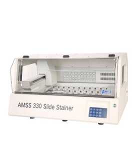
Slide Stainer
[a]https%3A%2F%2Fwww.amos-scientific.com%2Fproducts%2Fslide-stainer%2F[/a] <br>#staining #machine #for #histology <br>The AMOS Slide Stainer is a highly advanced instrument designed for automated staining of histology slides. It offers precise and efficient staining protocols, ensuring consistent and reproducible results. The stainer accommodates multiple slides and provides a user-friendly interface for easy operation. The AMOS staining machine for histology is available for sale, providing a reliable and efficient solution for slide staining in research, diagnostics, and educational laboratories. <br> <br>The Various Staining Techniques Performed with Slide Stainers <br>1. Routine Hematoxylin and Eosin (H&E) Staining: H&E staining is the most commonly performed staining technique in histology and pathology. It involves the use of hematoxylin to stain cell nuclei blue-purple and eosin to stain cytoplasm pink. Slide stainers automate the process by precisely timing and controlling the staining steps, ensuring consistent and reliable results for the visualization of tissue structures. <br> <br>2. Immunohistochemistry (IHC): automatic stainer in histopathology are widely used for immunohistochemistry, a technique that allows the detection and visualization of specific proteins in tissue samples. The slide stainer automates the incubation steps with primary antibodies, secondary antibodies, and the chromogen, providing precise timing and temperature control. This ensures reproducibility and accurate protein detection in diagnostic and research applications. <br> <br>3. Special Stains: Slide stainers are capable of performing a variety of special stains that allow visualization of specific tissue components or abnormalities. Examples include silver staining for highlighting reticular fibers, Masson's trichrome staining for collagen visualization, or Periodic Acid-Schiff (PAS) staining for glycogen detection. The automation provided by slide stainers ensures consistent and standardized staining protocols for these special stains. <br> <br>4. Fluorescence In Situ Hybridization (FISH): Slide stainers equipped with fluorescence capabilities can perform FISH staining. This technique utilizes fluorescently labeled DNA probes to target specific DNA sequences in the tissue sections, allowing for the detection of genetic aberrations, such as gene amplifications or chromosomal rearrangements. Slide stainers automate the various hybridization and wash steps involved in FISH, ensuring reproducibility and accuracy in the detection of genetic changes. <br> <br>These staining techniques, performed with the assistance of slide stainers, are essential for visualizing specific tissue components, identifying pathological changes, and aiding in the diagnosis and research of various diseases. The automation provided by slide stainers improves efficiency, reproducibility, and standardization in the staining process, ultimately enhancing the quality and reliability of histological and pathological analysis. <br> <br>Advantages of Slide Stainers in Slide Staining Processes <br>1. Automation: Slide stainers automate the staining process, reducing the reliance on manual labor and minimizing the risk of human error. Automated protocols ensure consistent timing, temperature control, and reagent dispensing, leading to reproducible and reliable staining results. <br> <br>2. Time savings: Automated slide stainers significantly reduce the hands-on time required for slide staining. By streamlining the staining process, slide stainers allow laboratory staff to focus on other tasks, increasing overall laboratory productivity. Batch processing capabilities enable multiple slides to be stained simultaneously, further saving time. <br> <br>3. Staining consistency: Slide stainers ensure consistent and standardized staining protocols, eliminating variations that may occur with manual staining methods. Consistency in staining improves intra-laboratory reproducibility and allows for accurate comparison of slides over time or between different laboratories. <br> <br>4. Reagent efficiency: Slide stainers optimize reagent usage, reducing waste and cost. Automated dispensing of reagents minimizes overconsumption and contamination risks, ensuring that reagents are used efficiently without compromising staining quality. This is particularly important for expensive or limited quantity reagents utilized in specialized stainings. <br> <br>5. Workflow integration: Slide stainers can be integrated with laboratory information management systems (LIMS) or digital imaging systems, streamlining the workflow and improving data capture and traceability. Connected systems enable efficient tracking of slides, reagents, and staining protocols, promoting seamless integration with downstream processes for analysis and archiving. <br> <br>For automatic slide stainer in histopathology and histopathology laboratory equipment, contact us.
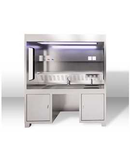
Pathology Grossing Stations
[a]https%3A%2F%2Fwww.amos-scientific.com%2Fproducts%2Fpathology-grossing-stations%2F[/a] <br>#pathology #grossing #stations <br>Pathology grossing workstation are essential equipment in histopathology labs. They are designed for the examination and dissection of surgical specimens, ensuring accurate and efficient tissue preparation. These stations often feature adjustable height settings, integrated ventilation, and ample workspace, providing a safe and ergonomic environment for pathologists. <br> <br>The Role of Pathology Grossing Stations in Histopathology Labs <br>histopathology grossing station play a critical role in histopathology labs by providing a controlled and efficient environment for gross examination and processing of tissue specimens. Here are four to five key roles of pathology grossing stations in histopathology labs: <br> <br>1. Tissue Examination and Dissection: pathology grossing stations provide a dedicated workspace for pathologists and histotechnologists to visually examine tissue specimens. They offer appropriate lighting, magnification, and ergonomic features to facilitate accurate assessment and dissection of tissues, ensuring representative sampling and precise identification of pathological features. <br> <br>2. Sample Preparation: grossing station for histopathology are equipped with specialized tools and accessories, such as cutting surfaces, forceps, and dissecting instruments, to assist in the dissection and processing of tissue specimens. They provide a standardized and controlled environment for precise and consistent specimen preparation, including proper orientation, trimming, and sampling for subsequent analysis. <br> <br>3. Containment of Biohazards: histopathology grossing instruments are designed with safety features to minimize the risk of exposure to biohazardous materials. They offer splash guards, ventilation systems, and containment areas to prevent the spread of infectious materials, protecting laboratory personnel and maintaining a safe working environment. <br> <br>4. Workflow Efficiency: Properly designed grossing stations are configured to optimize workflow efficiency in histopathology labs. They are often equipped with features such as integrated storage areas, dictation systems, and connectivity interfaces for seamless integration with laboratory information systems (LIS). This streamlines the grossing process, facilitates data entry and documentation, and enhances overall laboratory productivity. <br> <br>5. Compliance with Regulations and Best Practices: Pathology grossing stations are built to comply with regulatory standards and guidelines, such as those set by the Occupational Safety and Health Administration (OSHA) and the College of American Pathologists (CAP). They are designed to meet ergonomic requirements, improve safety, and ensure adherence to best practices in tissue handling and processing during gross examination. <br> <br> <br> <br>In summary, pathology grossing stations are indispensable in histopathology labs. They provide the necessary infrastructure, tools, and safety features to perform accurate tissue examination, standardized sample preparation, and efficient workflow management. By supporting the precise and controlled processing of tissue specimens, these stations contribute to the accuracy, reliability, and safety of histopathological diagnostic assessments. <br> <br>Safety Measures in Using Pathology Grossing Stations <br>1. Personal Protective Equipment (PPE): Always wear appropriate PPE, including gloves, lab coats, and protective eyewear, when working at a pathology grossing station. PPE helps prevent exposure to potentially hazardous substances and protects against contamination. <br> <br>2. Ventilation: Ensure that the grossing station is adequately ventilated to remove potentially harmful fumes or odors. Proper ventilation helps maintain a safe and clean working environment. <br> <br>3. Chemical Handling: Handle chemicals used in grossing stations with caution. Read and follow the safety data sheets (SDS) for each chemical, including fixatives and reagents, to understand their potential hazards, proper handling procedures, and potential interactions. <br> <br>4. Sharp Instruments: Utilize proper techniques when working with sharp instruments, such as scalpels or blades, to minimize the risk of injury. Ensure proper disposal of used sharps in designated containers. <br> <br>5. Containment and Splatter Prevention: Use a proper containment system, such as a containment hood or splash guards, to prevent splatter and aerosol generation during tissue processing. This helps minimize exposure to potential biohazards. <br> <br>6. Regular Cleaning and Disinfection: Maintain a clean and sanitized work area. Regularly clean and disinfect the grossing station and surrounding surfaces to prevent cross-contamination and ensure a safe working environment. <br> <br>7. Emergency Preparedness: Familiarize yourself with the location and proper use of emergency equipment, such as eyewash stations and fire extinguishers. Know the relevant emergency response protocols and procedures specific to your laboratory. <br> <br>8. Training and Education: Ensure that you receive proper training and education on the operation and safety guidelines specific to the pathology grossing station. Stay updated on best practices and industry-standard safety protocols. <br> <br>Maintenance and Care for Pathology Grossing Stations <br>1. Regular Cleaning and Disinfection: Clean the grossing station regularly to prevent the accumulation of tissue debris, fluids, and contaminants. Use appropriate cleaning agents and disinfectants recommended by the manufacturer to maintain a sterile and hygienic working environment. Pay particular attention to areas that come into direct contact with specimens or body fluids. <br> <br>2. Check Ventilation System: The ventilation system plays a vital role in removing hazardous fumes and maintaining a safe working environment. Regularly inspect and clean ventilation filters, fans, and ducts to ensure proper airflow. Consult the manufacturer's guidelines for specific maintenance instructions on the ventilation system. <br> <br>3. Calibration and Inspection: Periodically check and calibrate components of the grossing station, such as the digital scales or electronic controls, to ensure accurate measurements and optimal performance. Inspect all parts, including cutting surfaces and movable components, for any signs of wear, damage, or malfunction. Follow the manufacturer's instructions for calibration and inspections. <br> <br>4. Sharps Disposal: Properly dispose of sharps used in the grossing station. Use designated sharps containers and ensure they are securely closed and regularly emptied following biohazard waste management guidelines. Safely handle and dispose of sharps to prevent injuries and potential contamination. <br> <br>5. Training and Maintenance Record Keeping: Train laboratory staff on proper usage and maintenance protocols for the pathology grossing station. Keep track of maintenance activities, inspections, and repairs in a maintenance log or record. This helps identify patterns, track equipment performance, and ensure that scheduled maintenance tasks are consistently implemented. <br> <br>For grossing table pathology and histopathology apparatus, contact us.
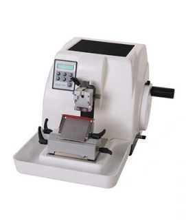
Microtome
[a]https%3A%2F%2Fwww.amos-scientific.com%2Fproducts%2Fmicrotome%2F[/a] <br>#microtome #for #sale <br>The AMOS Microtome is a precision instrument used for cutting thin and uniform sections of tissue samples. It offers precise control over section thickness, ensuring high-quality results for histology and pathology applications. The microtome in histology features adjustable cutting speeds, specimen orientation, and a range of accessories for various cutting techniques. The AMOS Microtome is available for sale, providing a reliable and efficient solution for tissue sectioning in research, diagnostics, and educational laboratories <br> <br>The Role of Microtomy in Research and Diagnostic Pathology <br>1. Histological analysis: microtome in histopathology is essential for the preparation of thin, uniform sections of tissue specimens for microscopic examination. These sections are then stained and mounted onto slides for histological analysis. Microtomy enables the visualization of cellular and tissue structures, aiding in the diagnosis and characterization of diseases in diagnostic pathology. It also facilitates research by providing crucial insights into the morphological features of tissues and cells. <br> <br>2. Immunohistochemistry (IHC): microtome for sale is often employed in immunohistochemistry, a technique used to detect and localize specific proteins or antigens in tissue sections. Thin sections prepared by microtomy allow for optimal antibody penetration, maximizing the sensitivity and accuracy of IHC analysis. By facilitating the visualization of protein expression patterns, microtomy helps researchers and pathologists identify disease markers, assess treatment response, and study various biological processes. <br> <br>3. Molecular pathology: Microtomy also plays a role in molecular pathology, where it is used for the preparation of tissue sections for molecular analyses, including DNA or RNA extraction, in situ hybridization, and fluorescence in situ hybridization. By obtaining well-preserved and properly sectioned tissue samples, microtomy ensures the accuracy and reliability of molecular studies, enabling genetic and genomic analysis, studying mutational profiles, and guiding personalized therapies. <br> <br>4. Tissue banking and research repositories: Microtomy is instrumental in the creation of tissue banks and research repositories. By cutting and mounting thin sections of tissue samples, microtomy facilitates their preservation and long-term storage. These preserved tissue sections can be utilized for future research studies, retrospective analysis, and validation of findings. The availability of well-preserved sectioned tissues enhances collaborative research and enables the sharing of valuable resources among pathologists and researchers. <br> <br>There are many histopathology equipment manufacturers, but we are one of the best choices for you.
About AMOS1 .com's Store
No description provided.
0.0
Customer Reviews
See what others are saying about AMOS1 .com's Store. We verify purchases to ensure reviews are genuine.
No reviews yet for this store.