

alexbrownss's Store
Owned by alexbrownss Joined May 2023 3 Listings
alexbrownss's Store
easyid.me/biz/alexbrownss-239dfa • Verified Store
Scan to open official EasyID digital store, view live catalog, or save business card.
Verified Merchant
EasyID Certified Seller
Fast 24h Packing
Direct courier dispatch
Zero Escrow Direct
Direct merchant settlement
Store Listings (3)

Phosphate pseudoUridine
Phosphate pseudoUridine is usually produced by the phosphorylation occurring in the 5’-position hydroxyl group of ribose of pseudoUridine. Phosphate pseudoUridine belongs to the C-nucleotides, in which the uracil moiety is combined with the sugar through its carbon atom rather than nitrogen atom forming a C-C glycosidic bond. The phosphate pseudoUridine can be divided in pseudoUridine 5ʹ-monophosphate (ΨMP), pseudoUridine 5ʹ-diphosphate (ΨDP) and pseudoUridine 5ʹ-triphosphate (ΨTP) depending on the number of linked phosphates. <br> <br>PseudoUridine 5ʹ-monophosphate (ΨMP) <br>PseudoUridine 5ʹ-monophosphate is an ester of phosphate and nucleoside and consists of a phosphate functional group, a pentose ribose and a base uridine. In theory, ΨMP can be obtained by twice hydrolysis of pseudoUridine 5’-triphosphate, or through pseudoUridine coupled with one phosphate. A study has corroborated a metabolic process of pseudoUridine, including the single phosphorylation of pseudoUridine to afford ΨMP catalyzed by pseudoUridine kinase, then the C-C glycosidic bond cleavage to produce uracil and ribose 5’-phosphate mediated by ΨMP glycosidase. However, the metabolism of PseudoUridine in eukaryotes has not been thoroughly studied. And the functions of ΨMP are also not supported by sufficient literature. Interestingly, one recent study showed that ΨMP is toxic to Arabidopsis thaliana, and ΨMP may cause delayed germination and growth inhibition of this plant. <br> <br>PseudoUridine 5ʹ-diphosphate (ΨDP) <br>PseudoUridine 5ʹ-diphosphate consists of two phosphate functional groups, a pentose ribose and a base uridine. The synthesis of ΨDP was reported as early as 1963, but to our knowledge, the biological applications of ΨDP are still to be discovered. The synthesis of PseudoUridine 5’-diphosphate has been achieved from PseudoUridine using the cyanoethylphosphate method to prepare the monophosphate and the nucleoside phosphoramidate method to make the diphosphate. During the synthesis, two side reactions occurred. One of these was an alkylation of the pyrimidine ring, the other involved an anomerization of the C-C glycosyl bond to give a steric isomer of ΨDP. <br> <br>PseudoUridine 5ʹ-triphosphate (ΨTP) <br>PseudoUridine 5ʹ-triphosphate consists of three phosphate functional groups, a pentose ribose and a base uridine. It is well known that nucleoside triphosphates (NTPs) are served as the building blocks for the synthesis of RNA molecules whether in vitro or in vivo, both mediated by the polymerase. In addition, the chemical synthesis of ΨTP can be achieved by the reaction of ΨMP with tributylammonium pyrophosphate under basic conditions. In recent years, the applications of mRNA in the field of cancer immunotherapy and other biomedical fields have received increasing attention from researchers. However, the biological therapy of mRNA is limited by the immune toxicity in the host. This issue has been improved via mRNA modification that utilizes the naturally occurring modified nucleotides, such as pseudoUridine-5’-triphosphate. This pseudoUridine modified mRNA can serves as a promising tool for gene replacement and vaccination. For mRNA modification, successful molecular biology applications depend heavily on the quality and purity of NTPs. The simple and efficient synthesis of ΨTP to afford a high purity product can support the modification of RNA.
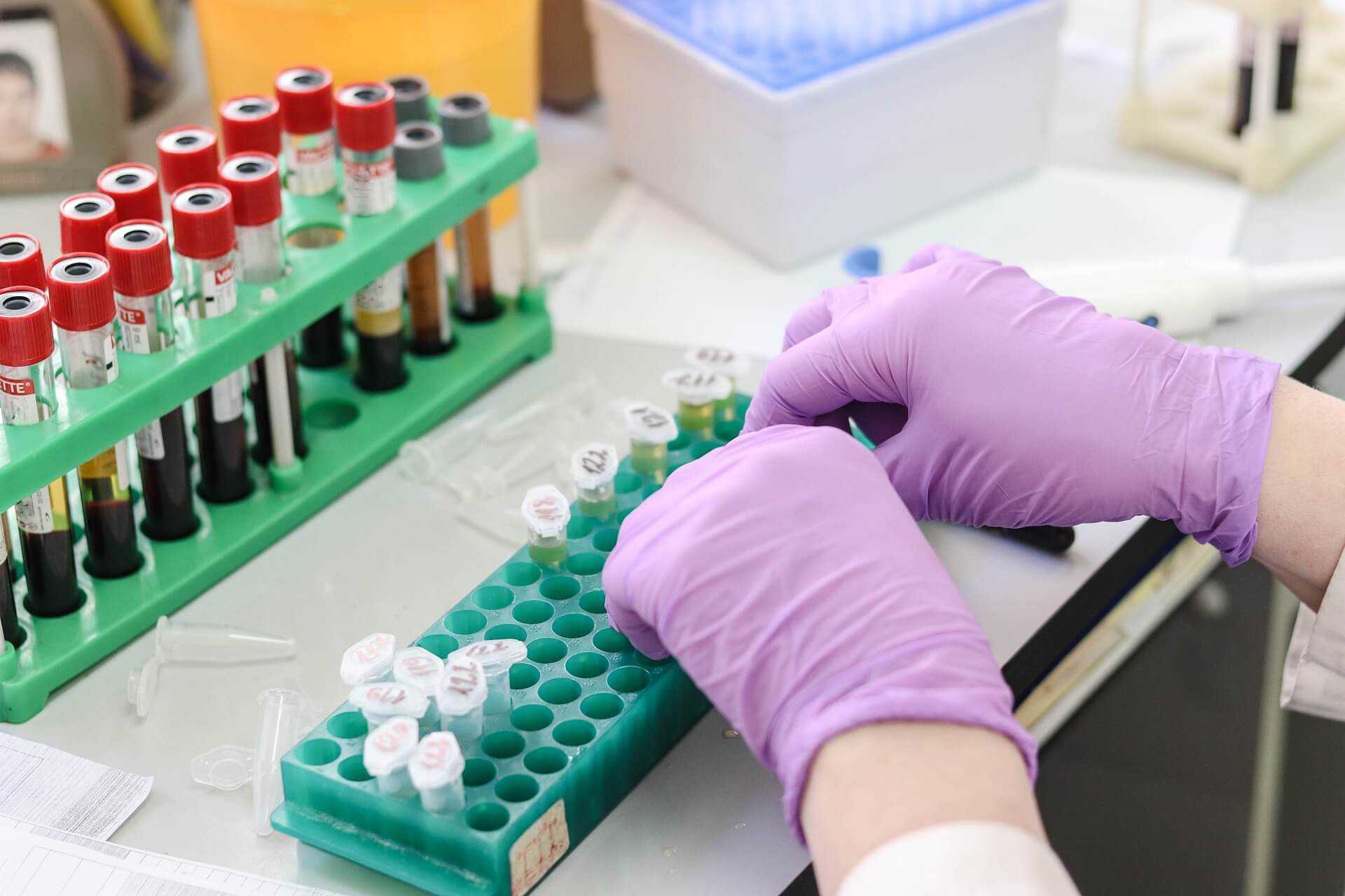
Antibody-Antibiotic Conjugation
BOC Sciences is a mature provider of antibody and ADC related services. In the past decade, we have accumulated a wealth of experience in antibody engineering, linker design, and pharmacological evaluation. Now, BOC Sciences's scientific team has been supporting customers at the forefront of antibody antibiotic conjugation development and will apply first-hand expertise to your unique project needs. <br> <br> <br> <br>Introduction <br> <br>Antibodies are an effective part of the immune system. For bacterial infections, antibodies provide omnidirectional protection to host cells at almost every stage of the pathogenesis. Studies have found that an antibody-antibiotic conjugate (AAC), which combines the key properties of antibodies and antibiotics, has been shown to be effective against Staphylococcus aureus infection. Preclinical trials showed that AAC was much better in scavenging Staphylococcus aureus in infected mice than standard vancomycin (antibiotics). AAC has three components: the bactericidal antibiotic payload, the antibody that targets the delivery of the payload to the bacteria, and the linker that attaches the payload to the antibody. The three components in AAC are all playing pivotal roles in determining overall selectivity and specificity, degree of safety and mode of action. <br> <br> <br> <br>Application <br> <br>In the face of rising antibiotic resistance and innovative gaps in the discovery of new antibiotics, the development of AAC provides an exciting opportunity for new treatments for infectious diseases by combining the pharmacological properties of antibodies and antibiotics into a single molecule. AAC may provide effective antibacterial compounds that may not be suitable as unbound drugs, such as poor pharmacokinetics or low therapeutic index. Finally, where the poor efficacy of antibiotics and disease recurrence are related to the ability of a variety of bacterial pathogens to survive in host cells, AAC can be used to improve the efficacy of antibiotics against these infectious diseases. <br> <br> <br> <br>Our Advantages <br> <br>Performed by trained professionals <br> <br>State-of-the-art analytical equipment <br> <br>Evidence of successful conjugation provided <br> <br>Our well-established drug discovery and antibody engineering platforms <br> <br>High levels of antibody and antibiotic recovery so you save precious reagents <br> <br>Strict quality control process to assure customers' complete satisfaction.
An Overview of Membrane-Associated E3 Ubiquitin Ligases
E3 ubiquitin ligase based on the ubiquitination degradation pathway has potential application value in the study of protein structure, function, and distribution. For example, E3 is involved in PROTAC-related research to target the degradation of pathogenic proteins. In addition, some E3 ligases have been reported to be novel biomarkers for certain diseases such as COVID-19. These studies indicate that E3 ligase is closely related to the occurrence and development of cancer. <br> <br> <br> <br>E3 ligase is distributed in many organs, cells, and subcellular compartments. Compared with E3 ligases distributed in other sub-cells, membrane-associated E3 ligases are more important in membrane organelles and play a crucial role in maintaining membrane morphology and function. <br> <br> <br> <br>E3 ubiquitin ligase located on the endoplasmic reticulum <br> <br>The endoplasmic reticulum (ER) is the transport system of eukaryotic cells and its main function is to synthesize proteins and lipids. Studies have shown that endoplasmic reticulum stress is closely related to the occurrence of cancer. Because of its significance in the metabolism of the endoplasmic reticulum, E3 ligase distributed in ER is crucial for the development of cancer. Gp78, TRIM13, and PARK2 are common E3 ligases distributed in ER that mediate carcinogenic effects through many different pathways, including cell proliferation, apoptosis, tumor angiogenesis, cell cycle disruption, cell metastasis, and mitochondrial function. <br> <br>- GP78 <br> <br> <br> <br>Gp78, also known as the autocrine motility factor receptor (AMFR), was originally found in B16-F1 melanoma cells. Gp78 has been found to promote tumor metastasis and migration. The ligand of Gp78 is AMF, and Gp78 is activated in response to the stimulation of AMF and then alters cell adhesion, proliferation, motivity, and apoptotic activity by activating downstream signaling pathways. In addition to being a receptor, Gp78 can also act as an E3 ligase to ubiquitinate and degrade some proteins. Therefore, Gp78 can work in the proteasomal degradation of endoplasmic reticulum-associated proteins. <br> <br> <br> <br>-TRIM13 <br> <br> <br> <br>TRIM13 is a tumor suppressor gene whose deletion is common in many malignancies. Its mRNA and protein expression is reduced in non-small cell lung cancer (NSCLC) tissues and cell lines. TRIM13 mainly distributes in the ER membrane, mediates the degradation of ER-related proteins, and regulates autophagy induced by ER stress. So far, caspase-8, MDM2, Akt, and Nur77 have all been identified as TRIM13 substrates, and some findings reveal that these substrates are involved in tumor cell apoptosis and proliferation. TRIM13 can induce ubiquitination of caspase-8, leading it to its transport to the autophagosome and activation, thus inducing cell apoptosis. In addition to caspase-8, TRIM13 forms a complex with MDM2 that negatively regulates tumor suppressor p53 and AKT to induce apoptosis. As a result of their interaction, MDM2 and AKT are ubiquitinated and degraded by the proteasome to increase the stability of p53 and reduce AKT kinase activity, thereby inducing apoptosis. <br> <br> <br> <br>-PARK2 <br> <br> <br> <br>PARK2 is a tumor suppressor gene related to microtubule stability, cell cycle disruption, mitochondrial homeostasis, cell apoptosis, and regulation of cell state metabolism. Current studies have confirmed the loss of PARK2 function in various human cancers, suggesting that its inactivation may contribute to tumor transformation and progression. In addition, PARK2 is involved in mitochondrial function regulation and transformation. By binding to mitochondrial DNA (mtDNA), PARK2 enhances TFAM-mediated mitochondrial transcription and restores PGC-1α expression, thus promoting mitochondrial production. PARK2 also regulates the activity of several apoptosis-related proteins in the Bcl-2 family, including Bax, Bcl-2, and Mcl1, promoting tumor cell apoptosis. <br> <br> <br> <br>-SYVN1 <br> <br> <br> <br>Studies have shown that SYVN1 can inhibit the growth and metastasis of breast cancer through the mir-96-5p/syvn1 axis. It interacts with IGF-1R to promote its ubiquitination and degradation, thus inhibiting the growth, migration, and invasion of breast cancer cells. In addition, SYVN1 can interact with heat shock protein 90 and promote ubiquitination of eEF2K to inhibit tumor cell proliferation. Some studies have shown that abnormal tumor cell metabolism may lead to ER damage and unfolded protein response (UPR). Typically, incorrectly folded proteins are monitored by chaperone proteins to be ubiquitinated by E3 ubiquitin ligase and degraded by the proteasome. Therefore, ER function is essential for cancer cell survival. <br> <br> <br> <br>E3 ubiquitin ligase located in the plasma membrane <br> <br>The plasma membrane not only regulates the flow of substances into and out of cells but also protects the integrity of cells and maintains their morphology. Numerous studies have demonstrated that frequent mutations of plasma membrane-associated E3 ligase can lead to cell carcinogenesis. RNF34, REFL, and NEURL1 are three E3 ligases distributed in the plasma membrane. They regulate carcinogenic effects through a variety of pathways, including cell growth and proliferation, migration, cell transfer, apoptosis, inflammatory, and immune responses. <br> <br>-RNF34 (CARP-1) <br> <br> <br> <br>Belonging to the CARP family, RNF34 can induce ubiquitination and degradation of death-effector domain (DED) caspase, thereby inhibiting the apoptosis of cancer cells. Moreover, RNF34 can inhibit the apoptosis of cancer cells by regulating the NOD1 pathway. NOD1 can not only induce apoptosis, but also control cell proliferation. Activation of NOD1 can lead to caspase-8 and caspase-9-induced apoptosis. In addition, RNF34 also stimulates the degradation of p53 and blocks the activation of reverse transcription, which means that it can also regulate the cancer cell cycle and accelerate cell growth and proliferation. <br> <br> <br> <br>-RFFL (CARP-2) <br> <br> <br> <br>RFFL, a member of the CARP-2 family, is an E3 ubiquitin ligase with a ring finger structure. Studies have shown that RFFL can inhibit the expression of p53 and promote the proliferation of cancer cells. In addition, RFFL plays a key role in cell migration. RFFL can induce ubiquitination and degradation of PRR5L, leading to MTORC2-mediated phosphorylation and activation of PKC, which can promote tumor cell migration. Therefore, RFFL may be a potential target to promote tumor migration and metastasis. <br> <br> <br> <br>-NEURL1 <br> <br> <br> <br>Neuralized-like 1 (NEURL1) is a tumor suppressor. At present, the subcellular localization of NEURL1 has not been determined. NEURL1 has been shown to significantly down-regulate protein levels through histone modification and has demonstrated a variety of tumor inhibitory functions, including promoting apoptosis and inhibiting tumor growth, angiogenesis, and invasion, mainly through the regulation of the Notch signaling cascade. In addition, NEURL1 can function as an E3 ubiquitin ligase by ubiquitinating the extracellular domain of membrane-bound protein Jagged1 and triggering the endocytosis and degradation of Jagged1. Reduced Jagged1 level will weaken the signal transduction between Jagged1 and the receptor Notch1 on the surface of adjacent cells. Inhibition of the Notch1 signaling pathway will down-regulate the transcription initiation of various cell cycle-related proteins, including HES1 and HEY1, thus inhibiting the growth and proliferation of tumor cells. <br> <br> <br> <br>-ZNRF3 <br> <br> <br> <br>Zinc And Ring Finger 3 (ZNRF3) is located on the plasma membrane and is often associated with the development of cancer. ZNRF3 regulates Wnt/β-Catenin signaling associated with cancer. Wnt stabilizes β-Catenin, which contributes to the formation of TCF/β-Catenin complexes and the regeneration of other coactivators to promote oncogene activation, such as c-MYC and cyclin D1, leading to the development of cancer. However, ZNRF3 can reduce the activity of the Wnt pathway and inhibit the proliferation of cancer cells by promoting the degradation of the Wnt receptor. <br> <br> <br> <br>-Cbl <br> <br> <br> <br>Cbl protein mainly exists on the surface of the cell membrane and plays an important role in tumorigenesis and anti-tumor immunity. Cbl expression has been reported to be associated with human colorectal cancer by inhibiting the migration of lung cancer cells and glioblastoma cells. Cbl-mediated ubiquitination can alter the cellular localization of proteins to regulate their function. First, it acts as an E3 ligase, activating the negative regulatory signal of RTK, and ubiquitinating and degrading RTK (receptor tyrosine kinase). Therefore, Cbl deficiency may lead to the development of malignant tumors and immune dysfunction. <br> <br> <br> <br>E3 ubiquitin ligase located in the Golgi apparatus <br> <br>The main function of the Golgi apparatus is to process, sort, and transport the proteins synthesized by the endoplasmic reticulum, and then send them to specific parts of the cell or secrete them outside the cell. The Golgi apparatus is particularly important for secreting proteins, and its important function of regulating metabolism mainly depends on the fluidity of the membrane and the abundance of anchor proteins. It has been reported that E3 ligase located in the Golgi apparatus is closely related to tumor hypoxia response and cell apoptosis. KLHL20 is an important Golgi-mediated E3 ligase that regulates cell carcinogenesis through a variety of pathways, including metabolic reprogramming, cell metastasis, tumor angiogenesis, and tumor growth. KLHL20 forms an E3 ligase complex with Cul3 and Roc1, which induces ubiquitination and degradation of substrate proteins such as PML and DAPK, thereby inhibiting apoptosis.
About alexbrownss's Store
No description provided.
0.0
Customer Reviews
See what others are saying about alexbrownss's Store. We verify purchases to ensure reviews are genuine.
No reviews yet for this store.